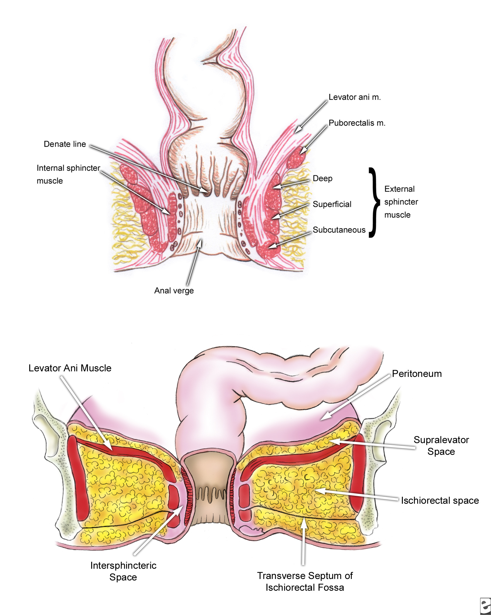
The Anal Canal
Definition
The last part of the GIT
Commences at the level where the rectum passes through the pelvic diaphragm
Ends at the anal verge
Parts
Internal sphincter : - Circular muscle fibres Thickened continuation of the circular muscle coat of the rectum - Pearly white
Spasm of this muscle is the cause of increased pain in many painful conditions of the anus.
Longitudinal muscle fibres - fan out to get attached to the skin of the anal verge.
The external sphincter
Posteriorly Attached to the coccyx
Anteriorly to midperineal point in the male and in the female fuse with the sphincter vaginae
Pink
The intersphincteric plane
The potential space between the internal and external sphincter
Contains 8-12 apocrine glands - can cause infections
Puborectalis Muscle:
This muscle maintains the angle between the rectum and the anal canal
The ano rectal ring : Junction between the rectum and the anal canal
The mucous membrane:
8-12 longitudinal folds known as the columns of Morgagni
Red
Anal valves are situated at the lower end of the columns of Morgagni
Just below the level of the anal valves transition of columnar epithelium to squamous epithelium - wavy margin - called the dentate line
The squamous epithelial lining of the anal canal is called the anoderm
Lower down the pale anoderm becomes pigmented
Arterial supply
By branches from the superior, middle and inferior haemorrhoidal arteries
Venous drainage
superior, middle and inferior haemorrhoidal veins
Lymphatic drainage
Hpper half to the para-aortic nodes
Lower half to the inguinal lymph nodes
Applied Anatomy
The submucosal venous plexuses engorge and form bleeding piles
Anal valves when get infected lead to perianal abscess